Chemical analysis with test strips
Introduction
The use of test strips for routine chemical analysis of urine is now commonplace. The strip consists of paper which contain absorbent material and reagents, fixed on a support. The color of the strip will change depending on the presence and concentration in the urine of specific substances and cells.
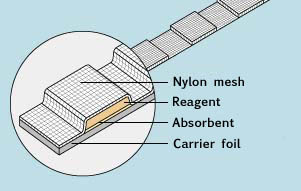
Test strip cross section.
Measurable constituents
Most diseases of the kidneys and urinary tract are accompanied by pathological changes in the urine composition. The most important changes such as bacteriuria, proteinuria, hematuria and leukocyturia can be detected quickly, easily and simultaneously. The following parameters can be evaluated:
- Specific gravity (density)
- pH
- Leukozytes
- Nitrite
- Protein
- Glucose
- Ketone bodies
- Urobilinogen
- Bilirubin
- Erythrocytes/Hemoglobin
Analysis with test strips can yield clinical information regarding kidney and urinary tract diseases, diabetic disorders, metabolic keto-acidotic disorders, liver diseases and hemolytic disorders.
The number of tests per strip may vary. The illustration below shows a color comparison scale.
Hint: urine samples may have their own strong coloring (in presence of bilirubin or extrinsic dyes, such as medicines), which can mask the true color of the reaction.
Complete urinalysis is the most frequently requested urinary test in hospitals and in physicians’ offices.
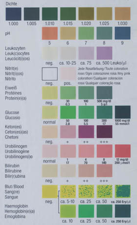
Colored scales of a commercial strip brand.
Procedure
The reaction can be interpreted visually or with an automatic reader.
 |
 |
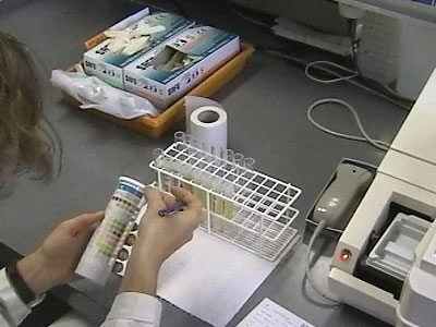
Visual interpretation |
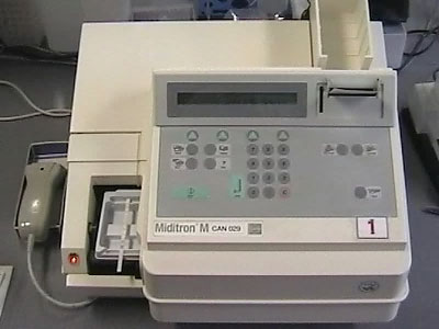
Automatic photometric test strip reader |
The sample must be as fresh as possible, mixed thoroughly but not centrifuged.
The tests are simple because each reactive zone contains a standardized amount of stable reagent:
- The test strip is immersed completely but briefly (maximum 1 second) in the sample. Then the one-minute countdown starts.
- Excess urine is removed by pressing the test strip against the test tube wall.
- The color of each reactive zone is compared visually with the colored scale printed on the package (the reading countdown can vary depending on the brand, use a chronometer). Color changes that appear on the edge of the strip or after 2 minutes are invalid and have no diagnostic value.
Interpretation by an automatic reader has the following advantages::
- It is not influenced by ambient light.
- The natural coloring of urine is accounted for.
- Individual and subjective variations in color interpretation are avoided.
Important
In order to avoid changes which might lead to incorrect results, urine specimens should not be stored more than 2 hours nor exposed to direct light.