Leukocytes
Clinical relevance
Leukocytes are found in urine in cases of inflammatory diseases of the kidney (pyelonephritis, interstitial nephritis), of the lower urinary tract (cystitis) or of the genital tract (vaginitis).
Identification
Neutrophils are the most frequent leukocytes found in the sediment. They are easily recognizable by their segmented nucleus and their granular cytoplasm. The cells have a diameter of 12-15 microns. Leukocytes are easily identified by Malbin Sternheimer staining. Fresh leukocytes are living cells, bright and pale blue in color (Sternheimer Malbin cells or bright cells). Their membrane is almost impermeable to dyes.
"Dark leukocytes" have lost this property. They are no longer alive. Typical nuclear segmentation is easier to recognize. In our opinion, systematic distinction between leukocytes from the kidney or leukocytes from the urinary tract is not possible by their color after staining.
“Dark cells” are most often observed in infections of the lower genito- urinary tract whereas "bright cells” observed in hypotonic urine often originate from diseases of the renal parenchyma. Since urinary tract infections often also involve renal parenchyma, both cell types can be found in the same urine.
Lymphocytes, monocytes, eosinophils and blasts (in case of myeloproliferative disorders) are rarely observed in urine.
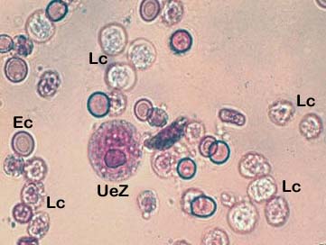
A round epithelial cell (UeZ) among numerous leukocytes and a few erythrocytes .
Terminology
- Leukocyturia: > 5 leukocytes/HPF (neutrophil polynuclear leukocytes).
A higher number indicates a moderate infection of the kidneys and/or urinary tract. - Pyuria: urinary sediment shows large amounts of leukocytes per field (over 50%) mostly of neutrophils, indicating an acute inflammatory process.
Differentiation
In some instances, neutrophils may be mistaken for renal epithelial cells. Renal epithelial cells have a larger, eccentric nucleus, and are often loaded with fat droplets.
Stability
Leukocytes are stable during 24h in high density, acidic urine. Lysis can occur within 3 hours in hypotonic and/or alkaline urine.