Renal cells
Renal epithelial cells are the only type of kidney cells. The term lipid cells will not be used (see below).
Renal epithelial cells
Clinical relevance
The most clinically relevant cells observed in urinary sediment are the renal epithelial cells (tubular).
Kidney disease, especially renal tubular damage, can lead to the abundant presence of renal epithelial cells in the urine. The normal urinary sediment may contain a small quantity of these cells, because of their constant renewal. Significant amounts of epithelial cells can also be encountered in cases of systemic viral infection such as cytomegalovirus or viral hepatitis, heavy metal poisoning (mercury, lead, uranium, cadmium etc.) because of tubular damage. Some cytostatic drugs (e.g. methotrexate, cisplatin) are also toxic to the kidney’s epithelial cells.
"Oval fat bodies" (degenerated epithelial cells) are often observed in nephrotic syndrome of any origin, as well as in advanced diabetes mellitus. The initial phase of renal transplant rejection is also often characterized by shedding of large quantities of renal epithelial cells.

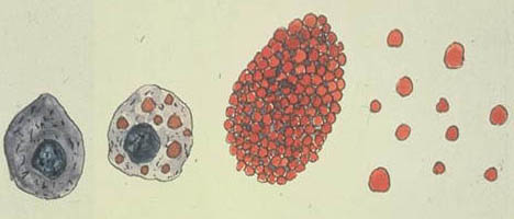
Schematic representation of renal epithelial cell degeneration: first without, then with lipid droplets; then transformation into oval fat bodies; and finally into free lipid droplets.
Terminology
These cells originate from the tubular epithelial layer. They usually are observed as isolated cells, but can also be observed in pairs or as casts.
Identification
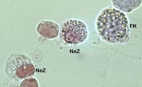
Renal epithelial cells without lipid droplets are difficult to differentiate from transition or round epithelial cells. They have almost the same size (20-30 μ m) as round epithelial cells (extra-renal cells). The kidney cells are polyhedral, and are not swollen as round epithelial cells. They have a round or oval shape with a slightly granular cytoplasm and a large round nucleus, often eccentric ("fried egg"). They are larger than leukocytes. When lipid droplets are present in the cytoplasm (birefringent bodies with polarized light microscope) their identification as epithelial cells is easy. A cell body filled with fat droplets and no visible nucleus is often referred to as an "oval fat body”. We avoid this term and only speak of kidney epithelial cells.

Renal epithelial cells (NeZ), from left to right: without lipid droplets; with lipid droplets; an oval fat body (FK) (brigjhtfield microscopy).
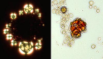
Fat droplets or free fat bodies can be found in urine or in casts (lipid casts). These fat bodies can have a diameter several times larger than epithelial cells. In polarized light microscopy, fat droplets display a "Maltese cross" shape (cholesterol ester). Staining the sediment with Sudan red is another method of lipid detection. The fat droplets are stained in red. Lipid birefringence should be distinguished from the artifact caused by starch grains that produce a pseudo Maltese cross image (see artifacts).

Fat body: polarized light (left), stained with Sudan red (right).