Extra-renal cells
Extra-renal cells are: round (transitional) and squamous epithelial cells, spermatozoa and neoplastic cells.
Round or transitional cells (urothelial)
Clinical relevance
Transitional epithelial cells are rarely observed in a routine examination of the sediment. They may originate from normal cell desquamation or from a urothelial carcinoma.
When a large number of transitional epithelial cells with nuclear abnormalities is observed, malignancy must be considered. In this case, a cytological examination is required.
Origin
The transitional epithelial cells originate from the epithelial lining of the renal pelvis, ureter, bladder, and of the 2/3 proximal urethral regions. Transitional epithelial cells are also called round epithelial cells.
Identification
The diameter of round epithelial cells is 20- 30μm. Their shape is round or oval with a relatively large, central, round nucleus. Their shape can vary depending on the amount of urine stored in the lower urinary tract, i.e. flat if the bladder is full and cubic if the bladder is empty. The physiological characteristic of these cells is that they absorb water, which gives them the appearance of a balloon. In the sediment, they appear singly or in pairs and are difficult to distinguish from renal epithelial cells.
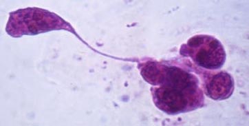
Round or transitional epithelial cells
Squamous epithelial cells
Clinical relevance
Epithelial cells have practically no clinical relevance, even when present in large amounts.
Origin
These cells originate from the distal part of urethra (male and female) and from the vagina.
Identification
These flat epithelial cells are larger (30-50μm in diameter) than all other cells found in the sediment. They are flat, round, oval, or polygonal and have a small, central, and condensed nucleus.
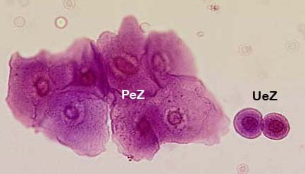
From left to right: squamous epithelial cells (PeZ); round cells (UeZ)
Spermatozoa
Spermatozoa are found frequently in urine from men. They can still be mobile. Some laboratories do not mention them in the result.
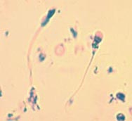
Spermatozoa
Neoplastic cells
Neoplastic cells are sometimes found in the urinary sediment in cases of malignant tumors of the lower urinary tract and prostate. Cytological techniques, requiring a larger amount of urine, are necessary to detect and identify these cells. Neoplastic cells are rarely detected in a urinary sediment sample because the small volume (10ml) required for a urinary status.
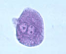
Neoplastic cell