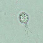
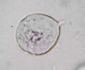
Trichomonas
They are found in the human oral cavity, intestines, vagina, and genital mucosa. The most frequent species encountered in the last two locations is Trichomonas vaginalis, a protozoan responsible for the majority of vaginitis (fluor vaginalis). It is found in the urethra in 75% of women and it is therefore not uncommon to find it in urine. Trichomonas are relatively large, they measure between 10 and 30 microns and are easy to recognize, when alive, due to the sawing movements of their flagella. They encapsulate frequently and are then difficult to distinguish from a renal epithelial cell. Living Trichomonas are stained pale blue with the Sternheimer-Malbin stain. The internal structure is hardly recognizable, but the flagella and undulating flagellar membrane are clearly visible in fresh urine.
Trichomonas belong to the class of protozoan flagellates. They have 4 anterior flagella and a recurrent flagellum raising a median undulating membrane attached to the cell body. As long as they are alive they are identified by the movement of their flagella and rapid and irregular motion of the cellular body. The oval nucleus is not visible and the cytoplasm is rich in granules.
The most frequent Trichomonas is Trichomonas vaginalis, causing trichomoniasis. Trichomonas vaginalis is pear-shaped and has a diameter of 7 -30 microns. It is transmitted through sexual intercourse. The urinary tract infection results in a pollakiuria and dysuria. In humans, it can infect the prostate, female vulva, vagina and, endocervical glands. Symptoms of Trichomonias vaginitis are abundant, watery, thick or foamy discharge and vaginal irritation and burning.
A trichomoniasis is frequently accompanied by a leukocyte exudation.
Trichomonas vaginalis